What SHBG Does to Hormone Availability in Perimenopause
High SHBG masks hormone deficiency while low SHBG creates unexpected androgen excess.
A standard hormone panel can say estrogen and testosterone are both "normal" while a woman feels the opposite of normal, and the reason often has nothing to do with how much hormone her body is making. It has to do with how much of it is actually free to act. Sex hormone binding globulin, or SHBG, is the protein that decides that question, and in perimenopause it moves in ways that make total hormone numbers close to meaningless on their own. This piece traces what SHBG does at the cellular level, how it shifts across the transition, and why a lab report without it is only telling half the story.
How SHBG controls hormone access at the cell level
SHBG is a glycoprotein made in the liver, and its job is to grab onto sex hormones (mainly testosterone and estradiol) and hold them in a form tissue can't touch. Think of it as a courier that picks up a package and simply never delivers it. The hormone is still in the bloodstream, and it still shows up on a total hormone test, but it is functionally inert as long as it's clamped to SHBG.
Circulating testosterone breaks into three fractions, and the math determines how much of it the body can actually use. Roughly 40% is bound tightly to SHBG, locked away from any tissue that might use it. About 2% floats completely unbound, free, and this tiny sliver is the most immediately active portion. The rest sits loosely bound to albumin, a much weaker attachment that lets the hormone detach and enter cells when needed, so it counts toward what's usable. The free fraction plus the albumin-bound fraction equals what's called bioavailable testosterone. The SHBG-bound 40% just doesn't count.
The free hormone hypothesis explains why total levels can be so deceiving. Research on SHBG has shown that when SHBG is experimentally elevated, total hormone concentration goes up (because more of it is bound and staying in circulation longer) while free testosterone stays flat, and the biological effect on target tissue actually weakens. In other words: more hormone on paper, less hormone doing anything in practice. That's the entire case for measuring free or bioavailable hormone instead of leaning on total values alone.
How SHBG shifts during the perimenopausal transition
Perimenopause typically starts sometime in the 40s and lasts an average of four years, though the range runs anywhere from two to ten. It is not a steady decline. Ovarian function tapers off, but hormone levels fluctuate along the way, meaning estrogen can surge before it eventually drops. The STRAW+10 staging system captures this by marking the early transition through cycle irregularity and the late transition through longer gaps between periods.
SHBG tracks this pattern because estrogen is one of its primary drivers. Estrogen binds to estrogen receptor alpha in the liver and upregulates SHBG synthesis directly, so when estrogen rises, as it often does in early perimenopause, SHBG rises right along with it. But what happens on the other side of that curve?
When estrogen eventually falls, later in the transition, SHBG falls too. Less SHBG means more free testosterone in circulation, even if the total testosterone number hasn't budged. That shift can produce a relative androgen excess, showing up as acne, new facial hair, scalp thinning, or fat that starts collecting around the midsection. None of this requires testosterone production to have changed. It only requires the protein holding it hostage to have backed off.
The metabolic factors that push SHBG further out of range in midlife women
Estrogen isn't the only lever on SHBG. Insulin might be the more dominant one in midlife, and it works in the opposite direction: high insulin directly suppresses SHBG synthesis in the liver. As estrogen declines across perimenopause, insulin sensitivity tends to worsen too. A woman can be dealing with low SHBG from two separate causes stacking on top of each other, the estrogen decline and the metabolic shift, at the same time.
The mechanism goes a step further than just suppression. Excess insulin also stimulates more frequent LH pulsatility, which drives more androgen production from the ovary, while simultaneously pushing SHBG down. That's a double effect: more androgen made, less protein to bind it, so free androgen availability climbs from two directions at once. Research has connected hepatic fat accumulation to lower SHBG in peri- and postmenopausal women, tying this back to a very concrete organ-level process rather than an abstract hormonal trend.
Body composition plays its own role separately. A 1997 cross-sectional study of 562 women found SHBG negatively correlated with BMI (r = −0.27), meaning higher body mass tracked with lower SHBG across the sample. Thyroid status also affects SHBG levels. Hyperthyroidism raises SHBG by stimulating hepatic synthesis, while thyroid underactivity can shift it in the opposite direction. Because thyroid-driven shifts can look identical to metabolic or estrogen-driven shifts on paper, thyroid function should get checked before anyone attributes an odd SHBG reading to menopause alone or to insulin resistance alone.
What SHBG does to usable estrogen and testosterone, and the symptoms that follow
High SHBG creates what amounts to a functional hormone deficiency. Total estrogen and testosterone can sit right in range, but with more of both bound and inactive, the felt experience mimics low hormone states across the board: low libido, fatigue, depression, mood disorders that seem to come from nowhere. Vaginal dryness occurs, along with decreased muscle and bone mass, irregular or absent periods, trouble building muscle in the gym, dry skin, thinning hair. A woman living through this can be told, correctly, that her total estradiol and total testosterone are normal, and still be entirely right that something is off.
Low SHBG produces close to the opposite picture, an androgen-excess state where more free testosterone is circulating and doing things it wasn't doing before. Acne returns in a woman's 40s the way it hasn't since adolescence. Oily skin, new hair growth on the face or body, hair loss from the scalp instead. Cycles get irregular, fat starts storing centrally, periods can turn heavier, and breast tenderness and mood swings appear. These symptoms get misattributed constantly, sometimes to stress, sometimes to thyroid issues, sometimes dismissed outright because the total testosterone reading looks unremarkable.
What makes perimenopause especially disorienting is the interaction between these two hormones through the same binding protein. When estrogen rises, it tells the liver to make more SHBG, and that SHBG binds more testosterone, so a perimenopausal estrogen surge can quietly reduce available testosterone even while total testosterone stays exactly where it was. Then estrogen drops, SHBG drops with it, and testosterone becomes more available again, sometimes abruptly. That single mechanism, estrogen driving SHBG driving testosterone availability, explains a fair amount of the unpredictability women describe during this stretch of years. One week libido is gone and energy is flat; a few months later it's acne and irritability. Same total numbers, different SHBG, different picture.
This also affects how hormone therapy performs. The same dose, the same formulation, can produce meaningfully different results in two women depending on where their SHBG sits. High SHBG can blunt the therapeutic effect of HRT before it ever reaches tissue, which goes a long way toward explaining why a protocol that works well for one woman barely moves the needle for another on paper-identical treatment.
Why oral versus transdermal estrogen produces dramatically different SHBG outcomes
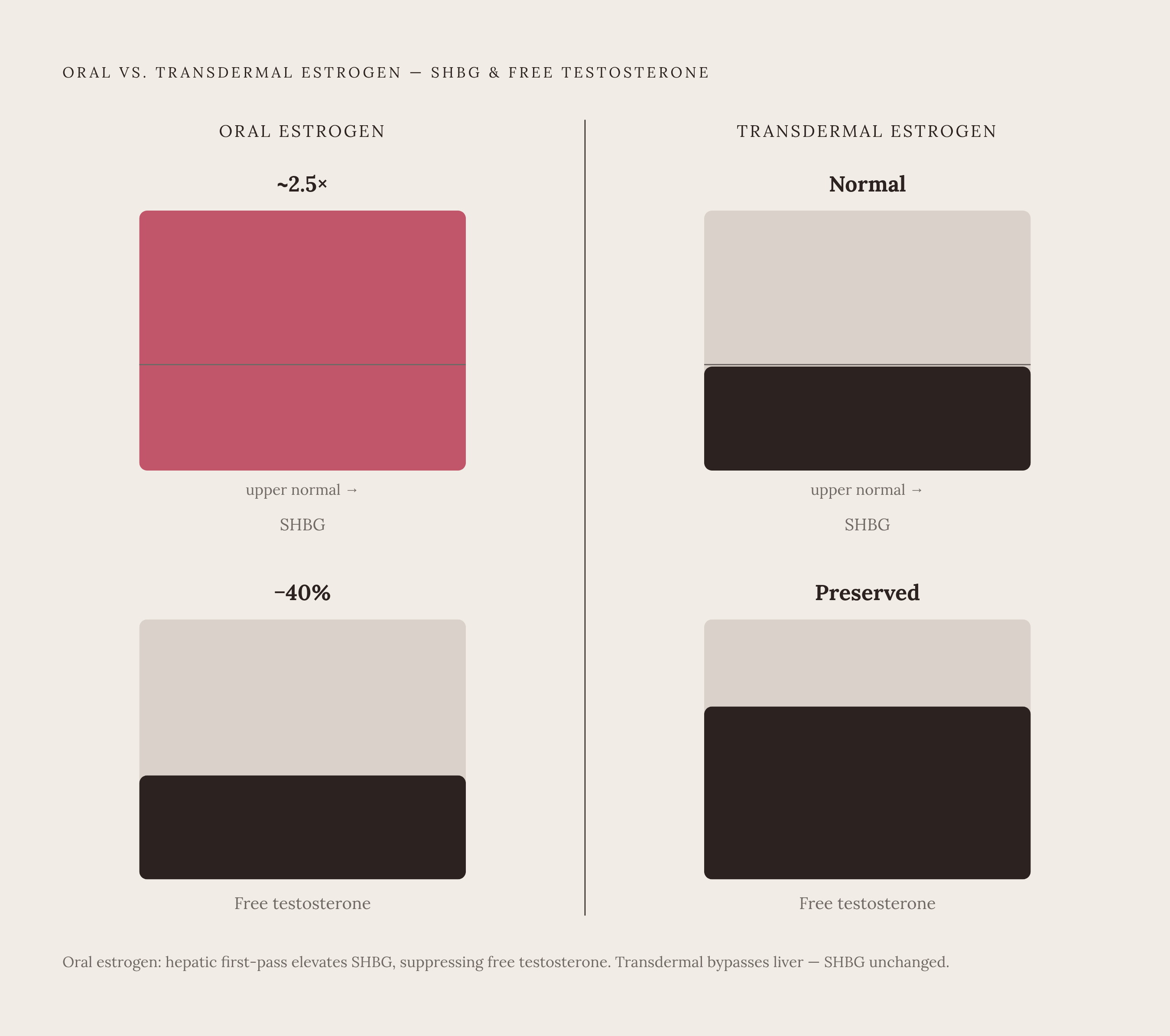
Route of administration affects SHBG because it determines where the estrogen goes first. Oral estrogen passes through the liver before it ever reaches the rest of the body, a process called first-pass metabolism. That hepatic exposure directly stimulates the liver to ramp up SHBG production. Transdermal estrogen, delivered through the skin, bypasses the liver on its way into circulation and largely skips this stimulation.
The difference in free hormone profiles between the two women is not subtle. In studies comparing delivery routes, the mean SHBG level in the oral estrogen group ran roughly 2.5 times higher than in groups not on oral estrogen, and pushed past the upper limit of the normal reference range. That's not a marginal shift, it's SHBG being pushed into territory that would be flagged as abnormal on its own.
The consequence for testosterone is where this becomes clinically relevant rather than just a lab curiosity. Free serum testosterone in the oral estrogen group measured about 40% lower than in the groups not taking oral estrogen. So oral estrogen, in raising estradiol, can end up paradoxically suppressing the very testosterone availability that governs libido and energy, working against the symptoms a woman may have started treatment to fix. Meanwhile, SHBG levels in the transdermal group looked comparable to women who received no estrogen replacement at all, which confirms something specific: it's the route of delivery driving the SHBG rise, not the estrogen molecule itself.
Why measuring total hormones without SHBG produces an incomplete and sometimes misleading picture
Most standard lab panels report total testosterone and total estradiol, full stop. They don't automatically report free or bioavailable levels, the fractions that actually determine what reaches tissue and produces an effect. That's the gap at the center of this whole discussion, and it's a structural gap, not a rare oversight.
Calculating bioavailable testosterone (free plus albumin-bound) isn't something most labs measure directly. It gets derived through a calculation that takes total testosterone, SHBG, and albumin as inputs. SHBG isn't a nice-to-have in that formula, it's load-bearing. Without it, calculating what's actually bioavailable becomes impossible.
The reference range for SHBG in women runs from roughly 14 to 144 nmol/L, a tenfold spread from bottom to top. A woman at 20 nmol/L and a woman at 100 nmol/L both fall inside "normal," yet their free hormone profiles from an identical total hormone reading look nothing alike. During the reproductive years, SHBG typically runs between 18 and 86 nmol/L, averaging around 38 to 40. Perimenopause brings its own range, 20 to 75 nmol/L, but layers in the erratic estrogen swings described earlier. Postmenopause widens it again, 16 to 115 nmol/L. A single "normal" label is doing a lot of work across a genuinely wide biological spread.
What drives SHBG also changes as a woman moves through this transition. Before menopause, estradiol is the dominant force setting SHBG levels. After menopause, insulin, waist-to-hip ratio, and testosterone itself take over as the dominant drivers. So the same SHBG number, say 40 nmol/L, carries a different meaning depending on which side of the transition a woman is standing on and what's actually pushing that number around. A lab value in isolation, without knowing where someone sits in the transition and without pairing it against total hormone levels, doesn't tell a complete story. It tells a fragment, and fragments leave so many women holding a "normal" lab report next to symptoms that feel like anything but.
Sources
- SHBG Hormone Levels: How Diet, Lifestyle, and Supplements Influence This Overlooked Hormone Marker
- Sex Hormone Binding Globulin (SHBG) Explained - Functional Fueling Nutrition
- High or Low SHBG Levels in Women: Treatment & Symptoms
- Changes in Sex Hormone-Binding Globulin Plasma... : Menopause
- ncbi.nlm.nih.gov
- SHBG Blood Test: MedlinePlus Medical Test
- Why you should care about Sex Hormone Binding Globulin (SHBG)
- onlinelibrary.wiley.com